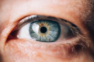

Age-related macular degeneration (AMD) is a leading cause of vision loss in individuals over the age of 50. An estimated 200 million people are affected worldwide; this number is expected to grow to 288 million by the year 2040.1 Because the eye is a direct extension of the brain and thus part of the central nervous system, AMD is the leading neurodegenerative condition in the world. Its prevalence far outnumbers cases of Alzheimer’s and Parkinson’s diseases combined.
AMD is a progressive condition that can significantly diminish vision if left unchecked. AMD preferentially affects the macula, the central part of the retina responsible for high resolution visual acuity. A healthy macula is critical for daily tasks, such as reading, driving, near work and recognizing faces. This disability caused by central vision loss from AMD can significantly impact an affected individual’s quality of life, independence and mental health, along with far-reaching socio-economic impact.
In this article, we will first review AMD with respect to its two types, stages, demographics and risk factors. Then, we will explore traditional ophthalmologic strategies for preventing and managing AMD, followed by integrative approaches that are based in nutrition, supplementation and lifestyle.
Understanding AMD
AMD is broadly categorized into two main types:
• Dry AMD: This is the more common form of macular degeneration, accounting for 85 to 90 percent of cases. In dry AMD, there is pathognomonic accumulation of drusen, which are yellowish-white, round deposits composed of extracellular lipids and proteins. “Drusen” is the German word for pebbles, and aptly, these deposits under the retina resemble small stones. Drusen are believed to be a result of oxidative stress and mitochondrial dysfunction. They often grow in size and number as macular degeneration progresses, triggering an inflammatory response.
• Wet AMD: Also, known as exudative or neovascular macular degeneration, wet AMD is less common, comprising about 10 percent of cases. However, wet AMD is more severe, accounting for 90 percent of cases of severe vision loss from AMD. In wet AMD, inflammation and the release of vascular endothelial growth factor (VEGF) lead to abnormal blood vessel growth under the retina. These tiny vessels leak fluid and blood, leading to distortion of vision and ultimately, rapid and painless central vision loss.
AMD can also be characterized by three distinct stages: early, intermediate and advanced. In the early stage of AMD, small drusen are present; however, vision typically is unaffected. In the intermediate stage, the drusen grow in size and number, and may coalesce to form larger lesions. In this stage, mild vision symptoms may occur, such as loss of contrast sensitivity with difficulty in dim lighting conditions.
The third stage—advanced AMD—may take the form of either geographic atrophy (GA) or neovascularization (nvAMD, aka wet AMD). In GA, there is significant thinning of the central retina with loss of retinal photoreceptors, while in nvAMD, there is leakage of blood, lipids, fluid and protein underneath or into the retina, leading to distortion of vision and/or central visual loss. Both types of advanced AMD may lead to legal blindness.
Whom Does AMD Affect?
AMD primarily impacts older individuals, with increasing prevalence with age. For example, in individuals between the ages of 50-59 years, the prevalence is estimated to be 2 percent. However, this number jumps to 30 percent in those above the age of 75 years.2 Women are also more affected than men, and a positive family history increases AMD risk.
In fact, genetics play a significant role in AMD. Over 50 identified genes have been linked to the condition. Short nucleotide polymorphism (SNP) gene variants, such as complement factor H (CFH) and age-related maculopathy susceptibility 2 (ARMS2), are associated with higher risk. Studies have shown that individuals with combinations of certain genetic markers are up to four times more likely to develop AMD compared to those without them.3 While the exact mechanism of many of the genetic variants for AMD are yet to be uncovered, genetic testing of 14 specific AMD SNP variants allows for personalized risk assessment over 10 years, along with targeted prevention strategies based on zinc sensitivity.4
Nutrition and lifestyle factors further exacerbate AMD risk, including:
• Smoking
• Low intake of the macular carotenoids, lutein and zeaxanthin
• High intake of processed and omega-6-rich foods
• Physical inactivity
• Ultraviolet (UV) light exposure
Air pollution has also been associated with increased risk of AMD, as demonstrated by a recently published meta-analysis. Higher levels of exposure to particulate matter less than 2.5 μm in diameter (PM2.5), nitrogen dioxide (NO2), and ozone (O3) were all associated with higher odds ratios of AMD.5
Conventional Treatment Modalities for AMD
The Age-Related Eye Disease Studies (AREDS and AREDS2) sponsored by the National Institutes of Health were landmark studies investigating the role of supplementation for AMD.6 The AREDS2 formula contains the following six ingredients: lutein (10 mg), zeaxanthin (2 mg), vitamin C (500 mg), vitamin E (400 IU), zinc (80 mg), and copper (2 mg). AREDS2 showed that supplementation with select nutrients reduced the risk of progression from intermediate to advanced AMD by 25 percent, and vision loss by 19 percent over five years. Unfortunately, for early dry AMD, supplementation was not effective in reversing or arresting disease.
For advanced AMD, there are several FDA (U.S. Food and Drug Administration)-approved treatments options, depending on whether the condition is wet or dry. For wet AMD (nvAMD), the standard of care involves injections into the eye (known as intravitreal injections) of anti-vascular endothelial growth factor (anti-VEGF) agents like bevacizumab, ranibizumab, aflibercept and others. These treatments target abnormal blood vessel growth and leakage, preserving vision in many cases. However, they are invasive, costly and require frequent administration every few months.
For advanced dry AMD (GA), there are two recently FDA-approved drugs on the market—pegcetacoplan and avacincaptad pegol. In clinical trials, both of these drugs (also given as injections into the eye dosed every few months) were demonstrated to slow lesion growth, but not improve visual acuity.
Preventive Strategies: An Integrative Perspective
Prevention is the cornerstone of AMD management, with integrative approaches that combine nutrition, lifestyle changes and environmental modifications.
Multiple studies have underscored the protective role of certain nutrients and dietary patterns, and the detrimental effects of others. Protective nutrients include the following:
• Macular Carotenoids (Lutein, Zeaxanthin and Meso-zeaxanthin): These yellow-pigmented macular carotenoids (also known as xanthophyll carotenoids) act as powerful antioxidants in the eye. They serve as our internal and natural UV and blue light filters. Lutein, zeaxanthin and meso-zeaxanthin are concentrated in the macula in a bull’s eye distribution, and are crucial for retinal health. Based on AMD research, the recommended daily intake of lutein and zeaxanthin is at least 6 mg, up to 20 mg, primarily from leafy green vegetables like kale, spinach, and collard greens, as well as eggs and corn.7 The highest quintile of intake of these macular carotenoids in population studies has been shown to reduce AMD risk by up to 40 percent.8
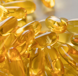
• Omega-3 Fatty Acids: Found in fatty fish, such as salmon, tuna, trout, and mackerel, the essential omega-3 fats, DHA and EPA, support retinal photoreceptor function and reduce inflammation. Regular consumption of omega-3-rich foods, equivalent to fatty fish two times per week, is associated with a lower risk of AMD progression.9
• Vitamins A, B6 and C, Folate, β-carotene, Lutein/Zeaxanthin, Magnesium, Copper: Long-term follow-up from AREDS has shown that these antioxidants and minerals have shown efficacy in reducing the risk of progression of AMD when obtained via diet.10 In another study, individuals who had several servings daily of fruits and vegetables rich in these antioxidants had a lower risk of AMD.9 These vitamins and minerals are also key ingredients in the AREDS2 formulation. However, the caveat is that in the context of certain genetic SNP combinations, excessive zinc (i.e., 80 mg, as found in the AREDS2 formulation) may double the risk of AMD.4
• Astaxanthin: An emerging nutrient, astaxanthin is a powerful red-pigmented carotenoid antioxidant derived from algae. Preliminary studies suggest that it is beneficial when taken as a supplement with the other macular carotenoids.11 Astaxanthin also has been shown to enhance macular health, improve blood flow and reduce oxidative stress.
Research has also highlighted certain foods that increase AMD risk. High-glycemic-index foods, such as white bread and sugary snacks, can exacerbate oxidative damage and inflammation. Similarly, diets high in saturated fats, processed meats and trans fats are linked to faster AMD progression.12 These findings emphasize the importance of avoiding these foods as an important dietary modification in the prevention of AMD.
A study published in JAMA Ophthalmology and endorsed by the American Academy of Ophthalmology (AAO) found that adherence to the Mediterranean diet lowers AMD risk by up to 43 percent.13 This diet emphasizes whole foods, including fruits, vegetables, whole grains, legumes, nuts, olive oil and moderate fish consumption. Reducing red meat and processed foods is also a key dietary strategy.
Beyond AREDS: Addressing Limitations
The AREDS and AREDS2 formulations are widely used for managing AMD. However, it is important to note they have several limitations: (1) these formulations were shown to be useful only for intermediate AMD, (2) the supplement formulations reduced progression by only 25 percent and (3) since these studies were published, additional nutrients have been identified that may help further reduce the risk of AMD.
These promising nutrients for AMD include:
• Meso-zeaxanthin: This lesser-known third macular carotenoid may trump lutein and zeaxanthin in the prevention of AMD, as it is concentrated at the very center of the macula.
• Astaxanthin: Despite its proven eye health benefits, it is missing from current AREDS formulations.
• Bioflavonoids: Compounds such as anthocyanidins, quercetin, resveratrol, rutin and hesperidin have been shown to be anti-inflammatory for eye disease, reduce oxidative stress and improve retinal blood flow.
• Sulforaphane: A phytochemical found in cruciferous vegetables, such as broccoli and broccoli sprouts, sulforaphane activates cellular detoxification pathways. The compound has been shown to support the retinal pigment epithelium, the cell layer underneath the retina in which drusen deposits form.
• Gamma-Linolenic Acid (GLA): An anti-inflammatory omega-6 fatty acid, GLA supports retinal health and may reduce inflammation.
In addition to targeted nutrition and supplementation, lifestyle and environmental exposures need to be considered in the management of AMD.
Smoking doubles the risk of AMD and accelerates its progression. Identical twin studies have highlighted the significant role of smoking as an environmental factor for AMD, even among individuals with a similar baseline genetic predisposition. Smoking induces higher levels of oxidative stress making smoking cessation programs a critical component of AMD prevention.14
Regular physical exercise should be encouraged for individuals at risk for AMD. Physical activity supports ocular health by improving blood flow and oxygenation, reducing inflammation and enhancing antioxidant defenses. Studies have shown that individuals who engage in moderate or high physical activity have a lower risk of AMD progression.15
Minimizing exposure to air pollution should also be a vital strategy in AMD prevention. The use of air purifiers and avoiding high-pollution areas can help mitigate risk.
Finally, emerging research has suggested that photobiomodulation using red and infrared light at specific wavelengths may help slow AMD and perhaps even improve visual acuity. Clinical trials are underway to establish safe practices to reap the benefits of light therapy while avoiding any potential damage to the eye’s delicate tissues.
Conclusion
Macular degeneration is an ocular and neurodegenerative condition that can lead to debilitating vision loss in older individuals. Both genetic and environmental risk factors are at play. Limited to no effective treatment options exist for intermediate and early stages of disease, respectively. Current FDA-approved treatments for advanced AMD include intravitreal injections; though effective at stabilizing disease in either nvAMD or GA, these injections must be administered every few months indefinitely and are costly.
Thus, prevention remains the most effective strategy for reducing overall AMD disease burden. Though AREDS and AREDS2 with their supplement formulations are considered the traditional standard of care for intermediate-stage AMD, research has shown that integrative approaches provide better protection against vision loss. The combination of strategic nutritional interventions, targeted supplementation beyond AREDS, and key lifestyle choices can empower patients to take control of their ocular health. By leveraging scientific evidence and incorporating insights from population data-based studies, practitioners can offer patients a proactive path to protect and preserve their vision.
To learn more about macular degeneration, download the free eBook, 5 Important Facts You Should Know About Macular Degeneration via this link: https://rudranibanikmd.activehosted.com/f/32. Also, stay tuned for my new book on macular degeneration, Beyond Leafy Greens – 7 Integrative Strategies to Prevent Macular Degeneration, set to release in 2025. To pre-order your copy, visit www.drranibanik.com/beyondleafygreens.
References:
1 Wong WL, Su X, Li X, Cheung CM, Klein R, Cheng CY, Wong TY. Global prevalence of age-related macular degeneration and disease burden projection for 2020 and 2040: a systematic review and meta-analysis. Lancet Glob Health. 2014 Feb;2(2):e106-16. doi: 10.1016/S2214-109X(13)70145-1. Epub 2014 Jan 3. PMID: 25104651.
2 National Institutes of Health, National Eye Institute. Prevalence of Blindness** Data–Data Tables, Summary of Eye Disease Prevalence Data: “Prevalence of Cataract, Age-Related Macular Degeneration, and Open-Angle Glaucoma Among Adults 40 Years and Older in the United States.”
3 Ross RJ, Verma V, Rosenberg KI, Chan CC, Tuo J. Genetic markers and biomarkers for age-related macular degeneration. Expert Rev Ophthalmol. 2007;2(3):443-457. doi:10.1586/17469899.2.3.443.
4 Vavvas DG, Small KW, Awh CC, Zanke BW, Tibshirani RJ, Kustra R. CFH and ARMS2 genetic risk determines progression to neovascular age-related macular degeneration after antioxidant and zinc supplementation. Proc Natl Acad Sci U S A. 2018;115(4):E696-E704. doi:10.1073/pnas.1718059115.
5 Wu J, Zhang Y, Xu X. Association between ambient air pollution and age-related macular degeneration: a meta-analysis. BMC Ophthalmol. 2024;24(1):202. Published 2024 Apr 30. doi:10.1186/s12886-024-03465-y.
6 Age-Related Eye Disease Study 2 Research Group. Lutein + zeaxanthin and omega-3 fatty acids for age-related macular degeneration: the Age-Related Eye Disease Study 2 (AREDS2) randomized clinical trial. JAMA. 2013 May 15;309(19):2005-15. doi: 10.1001/jama.2013.4997. Erratum in: JAMA. 2013 Jul 10;310(2):208. PMID: 23644932.
7 Seddon JM, Ajani UA, Sperduto RD, Hiller R, Blair N, Burton TC, Farber MD, Gragoudas ES, Haller J, Miller DT, et al. Dietary carotenoids, vitamins A, C, and E, and advanced age-related macular degeneration. Eye Disease Case-Control Study Group. JAMA. 1994 Nov 9;272(18):1413-20. Erratum in: JAMA 1995 Feb 22;273(8):622. PMID: 7933422.
8 Wu J, Cho E, Willett WC, Sastry SM, Schaumberg DA. Intakes of Lutein, Zeaxanthin, and Other Carotenoids and Age-Related Macular Degeneration During 2 Decades of Prospective Follow-up. JAMA Ophthalmol. 2015 Dec;133(12):1415-24. doi: 10.1001/jamaophthalmol.2015.3590. PMID: 26447482; PMCID: PMC5119484.
9 de Koning-Backus APM, Buitendijk GHS, Kiefte-de Jong JC, Colijn JM, Hofman A, Vingerling JR, Haverkort EB, Franco OH, Klaver CCW. Intake of Vegetables, Fruit, and Fish is Beneficial for Age-Related Macular Degeneration. Am J Ophthalmol. 2019 Feb;198:70-79. doi: 10.1016/j.ajo.2018.09.036. Epub 2018 Oct 10. PMID: 30312575.
10 Agrón E, Mares J, Clemons TE, et al. Dietary Nutrient Intake and Progression to Late Age-Related Macular Degeneration in the Age-Related Eye Disease Studies 1 and 2. Ophthalmology. 2021;128(3):425-442. doi:10.1016/j.ophtha.2020.08.018.
11 Piermarocchi S, Saviano S, Parisi V, Tedeschi M, Panozzo G, Scarpa G, Boschi G, Lo Giudice G; Carmis Study Group. Carotenoids in Age-related Maculopathy Italian Study (CARMIS): two-year results of a randomized study. Eur J Ophthalmol. 2012 Mar-Apr;22(2):216-25. doi: 10.5301/ejo.5000069. PMID: 22009916.
12 Seddon JM, Cote J, Rosner B. Progression of age-related macular degeneration: association with dietary fat, transunsaturated fat, nuts, and fish intake. Arch Ophthalmol. 2003 Dec;121(12):1728-37. doi: 10.1001/archopht.121.12.1728. Erratum in: Arch Ophthalmol. 2004 Mar;122(3):426. PMID: 14662593; PMCID: PMC8443211.
13 Merle BMJ, Colijn JM, Cougnard-Grégoire A, de Koning-Backus APM, Delyfer MN, Kiefte-de Jong JC, Meester-Smoor M, Féart C, Verzijden T, Samieri C, Franco OH, Korobelnik JF, Klaver CCW, Delcourt C; EYE-RISK Consortium. Mediterranean Diet and Incidence of Advanced Age-Related Macular Degeneration: The EYE-RISK Consortium. Ophthalmology. 2019 Mar;126(3):381-390. doi: 10.1016/j.ophtha.2018.08.006. Epub 2018 Aug 13. PMID: 30114418.
14 Mauschitz MM, Schmitz MT, Verzijden T, Schmid M, Thee EF, Colijn JM, Delcourt C, Cougnard-Grégoire A, Merle BMJ, Korobelnik JF, Gopinath B, Mitchell P, Elbaz H, Schuster AK, Wild PS, Brandl C, Stark KJ, Heid IM, Günther F, Peters A, Klaver CCW, Finger RP; European Eye Epidemiology (E3) Consortium. Physical Activity, Incidence, and Progression of Age-Related Macular Degeneration: A Multicohort Study. Am J Ophthalmol. 2022 Apr;236:99-106. doi: 10.1016/j.ajo.2021.10.008. Epub 2021 Oct 22. PMID: 34695401.
15 Velilla S, García-Medina JJ, García-Layana A, Dolz-Marco R, Pons-Vázquez S, Pinazo-Durán MD, Gómez-Ulla F, Arévalo JF, Díaz-Llopis M, Gallego-Pinazo R. Smoking and age-related macular degeneration: review and update. J Ophthalmol. 2013;2013:895147. doi: 10.1155/2013/895147. Epub 2013 Dec 4. PMID: 24368940; PMCID: PMC3866712.
Dr. Rudrani (Rani) Banik is a board-certified ophthalmologist and fellowship-trained neuro-ophthalmologist with additional certification in integrative and functional medicine. Dr. Banik focuses on the root cause of eye diseases, and uses integrative strategies for conditions, such as thyroid eye disease, macular degeneration, cataract, dry eye, glaucoma and other autoimmune diseases of the visual system. She runs a private practice based in New York, NY and is also associate professor of Mount Sinai in New York City where she serves as an educator and researcher. Dr. Banik is frequently featured as an expert in the media and has been interviewed on Good Morning America, CBS, NBC, ABC, The New York Times, The Washington Post and Fox, amongst many others. Dr. Banik’s first book, Beyond Carrots – Best Foods For Eye Health A to Z focuses on the 30-plus nutrients and 40 foods that best provide complete nutrition for your eyes. Its companion cookbook, The Beyond Carrots Cookbook, includes more than 160 delicious and nutritious recipes.